Radiation therapy is a pillar of oncologic care for various solid malignancies in the curative and palliative settings. Technologic advancements spanning over a century have now provided opportunities for radiation to be delivered to the target of interest with high accuracy and precision. In this regard, implementation of 3-dimensional (3D) conformal radiation therapy and intensity-modulated radiation therapy (IMRT) coupled with daily image guidance have enhanced the achievable therapeutic ratio over a variety of dosing and fractionation schemes. 1
Similar to medical oncology, radiation delivery has been guided by the fine balance of normal tissue toxicity and sustainability of durable tumor control. It is by these historical observations in which radiation dose has been chosen across a spectrum of malignancies and, to this day, remains the current dosing scheme for many cancers. Although it is generally accepted that tumors of the same stage, anatomic location and histology vary in their responses to radiation therapy, our field delivers treatment under a premise of established “clinical tolerance guidelines” rather than robust, tumor-specific, dose-response profiles.
In the last several decades, substantial advancements have been made in understanding the molecular catalog, metabolic networks and influence of the microenvironment on growth, spread and treatment response of various tumor types, yet employing these data in clinical decision-making has yet to inform the practice of radiation oncology. Furthermore, high-throughput analyses of clinically employed imaging modalities in radiation delivery has provided further opportunity to noninvasively categorize intrinsic tumor features and stratify patient outcomes. Further understanding of host and tumor differences with these interrogative approaches may provide the opportunity to precisely deliver radiation therapy beyond spatial and anatomic features, to one guided by intrinsic tumor biology.
Interrogation of Tumor Genomic Blueprints and Exploitation for Radiation Therapy
A major focus of personalized oncology has been the molecular characterization of tumors to identify unique druggable targets and generate higher order tumor classification methods to translate into clinical care. 2 Numerous high-throughput “-omics” analyses, which encompass transcriptional, proteomic, methylation, metabolomic and sequencing data, have provided unprecedented insight into the underlying biology of various human tumors. 3 These efforts have been largely performed within The Cancer Genome Atlas (TCGA) and International Cancer Genome Consortium (ICGC) programs, although several academic and commercial organizations now perform Clinical Laboratory Improvement Amendments (CLIA)-certified analyses of tumor tissue to complement these efforts. 4
The beginnings of precision oncology began with prior laboratory work, which identified the first cancer-related gene mutation in HRAS several decades ago. 5 Following this discovery, other somatic alterations have been identified in various tumors, which has formulated the notion that genetic alterations may be targeted in specific tumors. Notably, analysis of genomic data from patient tumors has provided opportunity to develop targeted agents against various proteins controlling kinases to epigenetic modulators. 6 Additionally, the development of therapeutic monoclonal antibodies (mAb) has transformed oncologic care, most recently by modulating the host immune response to tumors. 7 Many of these targeted agents have been employed in unselected metastatic cohorts, although tumor profiling has been able to separate responders from nonresponders based on intrinsic tumor features. Various trials employing molecular profiling have begun, though to date, no prospective trial has demonstrated a benefit to selecting targeted agents based on tumor genomic make-up. 8
In contrast to targeted therapy selection, determination of the optimal radiation regimen may require a different approach. Ionizing radiation does not have a distinct “target,” but distributes its effect in the cell via a stochastic manner causing damage to DNA, organelles and cellular membranes. 9 Additionally, at the tumor level, radioresponsiveness is influenced by other treatment parameters, including dose-volume relationships, total dose, fractionation pattern and type of radiation. In support of the latter, recent studies highlighted molecular differences in tumor cell radiosensitivity between dense and sparse ionizing rays 10 and various dose per fraction regimens. 11
Some of the first studies evaluating a molecular basis for radiation sensitivity were related to normal tissue toxicity in patients with alterations in ataxia telangiectasia mutated (ATM), 12 which supported a DNA damage basis for intrinsic radiosensitivity. Numerous observations in single nucleotide polymorphism (SNP) analyses and experimental manipulations of DNA damage repair (DDR) modulators have supported this model, yet no clinically actionable genetic alteration has been validated. 4 Interestingly, patients with rare genetic syndromes driven by compromised DDR pathways demonstrate a spectrum of responses, suggesting that a single alteration in core DDR machinery may not be a sole determinant of radioresponsiveness. 13 Yard et al profiled more than 500 cell lines and identified interconnectedness between DDR protein alterations and genomic stability, which governed intrinsic radiosensitivity. 14 This study underscores the polygenic trait of radiation sensitivity.
Attempts to model the polygenic nature of radiation sensitivity have continued to emerge in recent years. One of the first studies to address this question was by Eschrich et al, who identified a cancer-agnostic diverse gene network, which modeled the cellular survival following 2 Gy in 48 cancer cell lines. 15 This network was reduced to 10 hub genes, from which a multigene expression signature was derived, termed the radiosensitivity index (RSI). The RSI has predicted for clinical outcomes in various patient cohorts treated with radiation, 16 and recently Scott et al demonstrated that substitution of a tumor-specific RSI value for the alpha variable in the linear quadratic model derives an actionable tumor feature termed the genomically adjusted radiation dose (GARD), 17 which can stratify clinical outcomes in patients treated with radiation. 18,19
Others have hypothesized that tumor type-specific evaluation of radiation sensitivity may provide more robust classifiers compared to cancer-agnostic approaches, although some have suggested that despite heterogeneous sites of tumor origin, a common transcriptional program may regulate radiosensitivity. 20,21 Table 1 is a nonexhaustive list of gene signatures developed to infer radiosensitivity. For instance, in breast cancer, Speers et al derived a transcriptional signature based on survival after 2 Gy in breast cancer cell lines and a patient cohort that predicted for local control in patients treated with radiation, 22 and Tramm et al identified a 4-gene signature that predicted for postmastectomy radiation benefit. 23 Similarly, the postoperative radiotherapy outcome score (PORTOS), a 24-gene signature in prostate cancer, has been validated as a predictive tool for assessing distant metastasis risk following postprostatectomy radiation. 24
| Cancer Type | Number of Genes in Signature | Training Cohort(s) | Validation Cohort(s) | Reference(s) |
|---|---|---|---|---|
| Breast | 34 | 343 patients | 605 patients and additional matched 524 patients | 25 |
| Cancer Agnostic (NCI-60 cell line panel) | 474 refined to 10 by systems biology methods | 48 cell lines | 852- Breast 73- Pancreas 270 -GBM TCGA 60- NSCLC 92- HNSCC 14- Rectal 12- Esophagus 42- Melanoma | 15, 17, 109-113, 149 |
| Breast | 51 | 16 cell lines and 343 patients | 228 patients | 22 |
| Prostate | 24 | 196 patients | 330 patients | 24 |
| Breast | 4 | 191 patients | 112 patients | 23 |
| Breast | 248 | 168 patients | 139 patients | 107 |
| Cancer Agnostic (NCI-60 cell line panel) | 31 | 60 cell lines | 1045-TCGA Breast 463- TCGA GBM 263- Glioma | 99-101 |
| Head and Neck | 5 (miRNA) | 2 lymphoblastic cell lines with ATM alteration from single patient | 435-HNSCC TCGA | 102 |
| Head and Neck (HPV-) | 13 | 86- TCGA HNSCC 32 HNSCC cell lines 128- TCGA HNSCC (HPV-) | 44 HNSCC patients 63 HNSCC (HPV-) 5 HNSCC cell lines 59 cell lines (NCI-60) | 103 |
| Head and Neck (HPV-) | 7 | 130 patients | 121 patients | 104 |
| Esophageal | 41 | 152 patients | 31 patients | 26 |
| Gastric | 11 | 371- TCGA Gastric | 371 patients (cross-validated from training) | 108 |
| Soft Tissue Sarcoma | 26 | 253- TCGA Sarcoma | 101 patients (cross-validated from training) | 105 |
| Cervical | 7 | 25 patients | N/A | 106 |
Combining gene signatures representing distinct biological processes may also improve the robustness of clinical classifiers. For example, Cui et al developed independent radiosensitivity and antigen processing/presentation signatures in breast cancer cohorts and found that integration of these signatures improved outcome stratification. 25 Zhang et al also found that integrating a 31-gene signature with the RSI, both derived similarly from the NCI-60 cell line panel, improved predictive ability in esophageal cancer patients. 26
Interestingly, many signatures proposed to delineate intrinsic radiosensitivity show little overlap, if any, with regard to gene sets. Is this due to a broadly conserved transcriptional program resulting from genotoxic stress or is there redundancy in the information of gene signatures? Despite publication of various gene signatures representing diverse biologic processes (eg, hypoxia, epithelial-mesenchymal transition, cell proliferation), prior studies have identified similarities in the predictive ability of diverse gene sets in a single dataset for similar clinical endpoints. For instance, Fan et al found a high concordance for nonoverlapping gene signatures in breast cancer, suggesting a common biologic underpinning. 27
Few studies investigating relationships between gene signatures and clinical outcomes prove the specificity of the derived signature by testing against a negative control signature. A study by Venet et al found that gene signatures unrelated to cancer biology (ie, effect of postprandial laughter, skin fibroblast localization, social defeat in mouse brains) were associated with overall survival in a breast cohort and found that only 18 of 47 (40%) signatures from the literature had the ability to outperform random signatures of similar size. 28
Functional redundancy of many gene signatures argues that robust statistical methods, including random permutation of genes selected to represent signature modules, are needed to avoid spurious associations with clinical outcomes. 29 As the number of gene signatures continues to grow, it is important to interrogate the biology of individual genes composing the signature since sophisticated bioinformatics analyses can overcome real biologic differences and ultimately lead to no downstream utility. 30
There are several important limitations to consider when implementing genomic-based strategies in clinical medicine. A major concern is the use of single-biopsy-site, tumor-profiling data to infer overall tumor biology. Tumors have significant spatial and temporal heterogeneity, 31,32 often with opposing prognostic gene expression profiles or targetable mutations in different tumor regions. Although heterogeneity is evident, selection of many targeted therapies and clinically useful gene signatures is informed by single-region analyses, 33 suggesting that the calculated signal in the readout may represent central biology in the tumor. Tumor profiling adds an additional level of complexity compared to signatures derived from cell cultures due to heterogeneous cell populations contributing to tumor composition. Aran et al found that noncancerous cell populations contribute to gene expression profiles and following adjustment for tumor purity, variation in differentially expressed genes and pathway enrichments were lost; this study emphasizes the need to correct for tumor purity. 34
Another important feature to consider is the assumption that a snapshot of tumor biology derived from a single biopsy is representative of biology as treatment progresses. Myriad evidence demonstrates adaptive changes following exposure to various treatments. 35,36 For example, radiation has been shown to induce alternative splicing, 37 which has the potential to increase transcriptome diversity. Another example of adaptation is in prostate cancer cells exposed to enzalutamide, which results in differential expression of genes regulating inflammation and various metabolic processes. 38 Thus, assuming an iso-effect response to each fraction of radiation may not provide a complete picture of the dynamicity in a responding tumor. 39
Lastly, and of utmost importance, is the required external validation of derived signatures before adoption into clinical practice. Rigorous testing in prospective randomized clinical trials or prospectively collected retrospective analyses of previous phase III trials are required to demonstrate robustness of the signature outside of the training and nonprospectively collected validation cohorts. The utility of genomic-based approaches in radiation has lagged as none of the aforementioned signatures have withstood scrutiny of the protective regulatory barriers needed to safeguard patients from implementation in clinical decision-making.
Radiomics: A Noninvasive Means to Assess Tumor Biology
Routine medical imaging, including computed tomography (CT), magnetic resonance imaging (MRI) or positron emission tomography (PET), is paramount to the diagnosis, treatment and follow-up of cancer patients. 40 Radiation oncologists approach data supplied by these anatomical and functional images differently than diagnostic radiologists, in that images are used to plan dose distributions that cover gross disease or regions at risk for spread. Although qualitative assessment by radiologists provides useful diagnostic information, each image contains a plethora of features that may be used for precise radiation delivery and treatment selection.
Radiomics refers to high-throughput extraction of quantitative image features from standard-of-care images, such as CT, MRI and PET followed by relation to biologic or clinical endpoints. 41-43 This noninvasive process allows for the ability to describe tumor characteristics while accounting for spatial and temporal heterogeneity. 44,45 Radiomics has the capacity to detect medical imaging phenotypes that are reflective of tumor features at the cellular level, with a prime example being 18-fluorodeoxyglucose PET (FDG-PET) representing glucose uptake. 41 Also, data obtained from quantitative image analysis can identify novel tumor features that complement clinical or genetic characteristics, thus improving the understanding of tumor biology. 43 Radiomics has potential to be a powerful tool to personalize clinical-decision algorithms, and novel methods continue to emerge for utilization in radiation therapy.
The workflow of radiomics involves several steps, including image acquisition, segmentation of the regions of interest (ROI), extraction of descriptive features, predictive modeling, and validation; each of these steps pose unique challenges described further below. 42,45
Image Acquisition
The first step of radiomics involves acquiring standard-of-care images. Although lack of standardized imaging protocols across institutions does not significantly affect clinical utilization, these diverse protocols do impact extraction of quantitative features. Heterogeneous source data increase the probability for noise interference, calibration error and unfruitful analyses. For this reason, nonstandardized multi-institutional radiomics can pose major challenges. There have been recent attempts to address this issue through standardization of the imaging protocol, including the Quantitative Imaging Biomarkers Alliance (QIBA) and the Quantitative Imaging Network (QIN). 46,47
Segmentation of Regions of Interest
The next step is segmentation of ROIs to determine which pixels/voxels within the image are to be analyzed. This step has been called the most challenging and contentious component of radiomics, and the segmentation process varies greatly across studies. 42 The process can be conducted manually, which can introduce bias through user variability 48 or can potentially be semi- or fully automated with newer approaches. 49,50
Extraction of Descriptive Features
Radiomic features can be divided into spatial (static) and temporal (dynamic) features. Static features are derived from shape, volume, voxel intensity and texture, whereas dynamic features represent changes in kinetics with time-varying protocols. 51 Semantic features, commonly used in radiology to qualitatively describe images (eg, spiculation, cavitation, necrosis), can be time-consuming to capture and do not provide more granular data for statistical modeling. Ongoing efforts with machine-learning methods strive to increase inter-reader agreement, lower variance, and augment more rapid data acquisition for semantic features. 52 Agnostic features, which quantitatively describe heterogeneity within the ROI (eg, wavelets, textures, histogram characteristics) can provide statistical inter-relationships between voxels and reveal hidden patterns. These features can be calculated by various texture matrices (eg, gray-level co-occurrence, neighborhood gray tone difference matrix); for a more thorough description of feature calculation please see the recent article by Rizzo et al. 53
The feature extraction process is variable across institutions with recent attempts to address this issue. The Image Biomarker Standardization Initiative (IBSI) is an international collaboration that works to standardize extraction of image biomarkers. 54 Additionally, an open platform termed Computational Environment for Radiological Research (CERR) has been introduced to improve reproducibility, speed and clinical integration of radiomics research. 55,56 Other open-source software to extract features includes RaCaT and LIFEx. 57,58
Predictive Modeling and Validation
Following feature extraction, data interrogation via manual statistical analysis or machine learning, is conducted to test for relationships between features, clinical endpoints or other questions of interest in a training model. Model building from a small sample size relative to the number of features can result in reduced accuracy and risk of overfitting. This potentially may be obviated by predetermining subsets of features to analyze or removing highly correlated variables, yet there are notable statistical considerations when analyzing large datasets. 59,60 Model validation, both internal and external, is a necessity for radiomics studies. Ideally, a successful model will perform similarly in training and validation cohorts. Beyond the scope of this article, Park et al provide a useful guide to assess model performance in radiomics. 61 When constructing predictive models with multivariable analysis, guidelines from transparent reporting of a multivariable prediction model for individual prognosis or diagnosis (TRIPOD) can help maintain reproducibility and transparency. 62
Clinical Applications
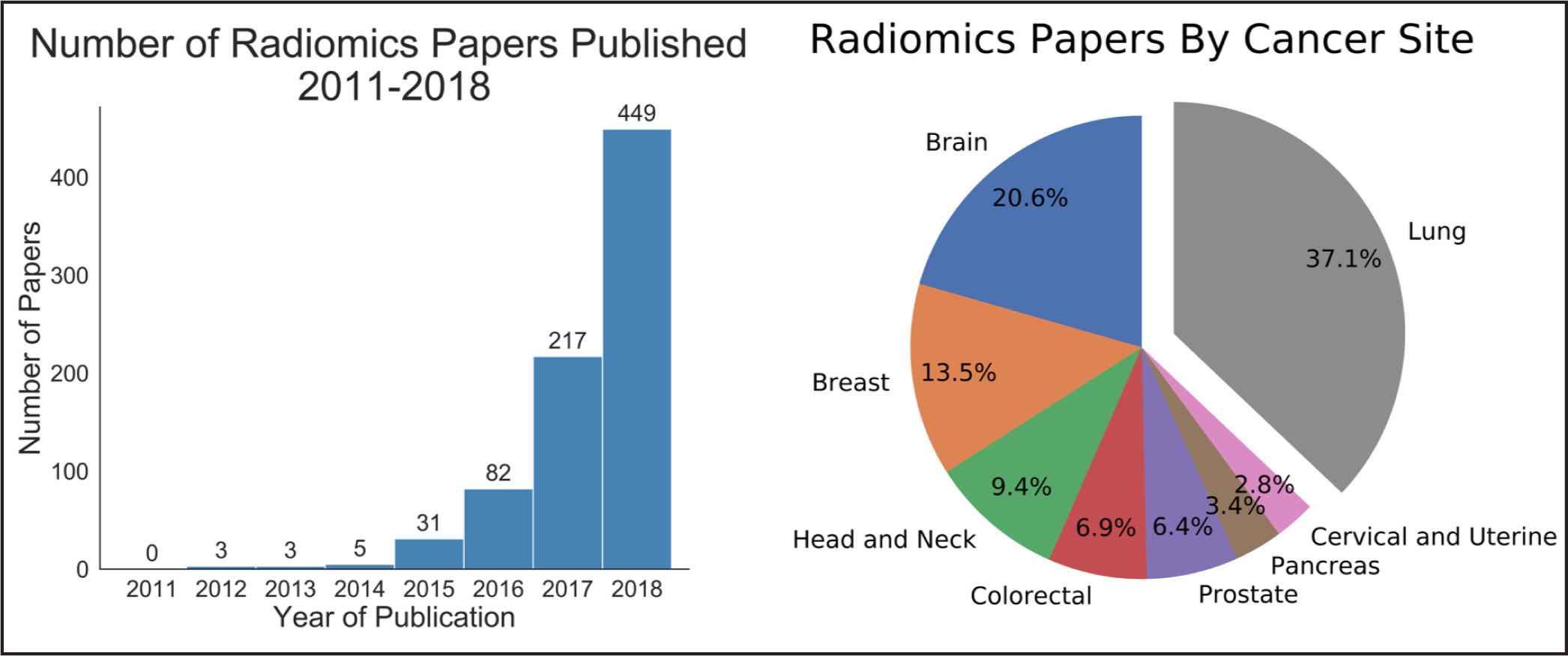
Radiomics can significantly impact clinical decision-making within oncology, 42,45,63 including radiation. 64 Due to the vast number of recent radiomics studies, indicated by a 50% increase in published studies between 2017 and 2018, 65 we have highlighted a subset with potential to personalize radiation therapy ( Table 2 ).
| Cancer | Imaging Modality | Study Endpoint(s) | Number of Patients | Conclusion of Analysis | Clinical Application | Reference |
|---|---|---|---|---|---|---|
| Prostate | MRI | Diagnosis | 381 | MRI-based radiomics models outperformed PI-RADSv2 in distinguishing cancerous vs non- cancerous tissue or high- vs low-grade disease | Diagnosis | 114 |
| NSCLC | PET | OS | Training: 262 Validation: 50 | FDG-PET radiomics from tumors and nodes can improve prognostication for NSCLC | Prognostication | 115 |
| GBM | MRI | OS | 79 | Multiregion quantitative analysis of MR images has prognostic utility for GBM and outperformed conventional prognostic factors | Prognostication | 74 |
| NSCLC | CT | OS, FFDM, LRC | 107 | Radiomics features change due to radiation therapy and end of treatment values may be indicators of treatment response | Prognostication | 116 |
| Prostate | MRI | Biochemical recurrence | 74 | Radiomic analysis of MRI predicted biochemical recurrence following radiotherapy | Prognostication | 117 |
| GBM | MRI | OS, PFS | Training: 126 Validation: 165” | Radiomic analysis had significant prognostic value for OS and PFS in patients with recurrent GBM receiving bevacizumab | Prognostication | 118 |
| Rectal | MRI | LR, DM, DFS | Training: 67 Validation: 34 | Delta radiomics via MRI predicted clinical outcomes after chemoRT and surgery as an independent prognostic factor | Prognostication | 119 |
| GBM | MRI | PFS, OS | 181 | Radiomics improved prognostication for patients beyond molecular, clinical, and standard imaging | Prognostication | 98 |
| GBM | MRI | OS | Training: 75 Validation: 37 | Deep-learning-based radiomics model was able to generate a prognostic imaging feature-based biomarker for OS prediction | Prognostication | 120 |
| NSCLC | CT | OS, RFS, LR-RFS | 59 | CT-based radiomics prognosticates OS and progression as early as 3 months after SBRT | Prognostication | 121 |
| NSCLC | PET/CT | OS, DSS, RC | 150 | Radiomics predicts control and survival for patients with lung cancer treated with SBRT | Prognostication | 122 |
| Head and Neck | PET/CT | LRC, DM | 300 | Models combining radiomic and clinical variables had significant prognostic utility for LRR and DM in patients treated with chemoRT | Prognostication | 123 |
| NSCLC | CT | OS | Training: 132 Validation: 62 and 94 | Subset of radiomic features from CT and CBCT images are interchangeable and a previously described radiomics signature is prognostic for OS | Prognostication | 66 |
| NSCLC | PET/CT | DM | Training: 70 Validation: 31 | PET imaging characteristics were significantly prognostic for the development of distant metastasis in patients with early stage NSCLC | Prognostication | 75 |
| Esophageal | CT | OS | 36 | Post-treatment texture analysis was predictive of survival, and the combination of pretreatment texture parameters and maximum wall thickness performed better than morphologic tumor response | Prognostication | 124 |
| GBM | MRI | OS | 32 | MRI spatial variations defined regional habitats in GBMs, and the distribution of these varied significantly among the different survival groups | Prognostication | 125 |
| Cervical | PET, MRI | LRC | Training: 69 Validation:33 | Radiomics from MRI and PET predicted recurrence and LRC with higher prognostic power than clinical parameters | Prognostication | 126 |
| NSCLC | CT | Molecular discrimination OS | 57 | Radiomic features from preoperative CT images were significantly associated with mutational profiles in lung squamous cell carcinoma | Prognostication Radiogenomics | 127 |
| Head and Neck | CT | Molecular discrimination discrimination LC | Training: 93 Validation: 56 | Heterogeneity of HNSCC tumor density is associated with LC after chemoRT and HPV status | Prognostication Radiogenomics | 95 |
| Prostate | CT | Gleason score | 342 | CT-based radiomics model was able to accurately distinguish high risk from low risk and Gleason score >7 vs 3+4 vs 4+3 | Prognostication Radiogenomics | 128 |
| Head and Neck NSCLC | PET/CT | Molecular discrimination OS | Training: 474 Validation: 545 | PET/CT-based radiomic signature was significantly prognostic for OS; radiomic features significantly associated with different gene sets | Prognostication Radiogenomics | 41 |
| Nasopharyngeal | MRI | Therapy response | Training: 100 Validation: 23 | MRI radiomics predicted response and survival and in combination with clinical data, showed excellent predictive performance | Prognostication Treatment Response | 129 |
| Hepatocellular | CT | LR | 106 | A robust radiomic signature (one signal feature) predicted LR and OS after radiation | Prognostication Treatment Response | 130 |
| Colorectal | CT | Molecular discrimination OS, PFS | 64 | Combining contrast-enhanced CT radiomics with gene expression and histopathologic factors provided improved prognostication | Radiogenomics | 97 |
| Head and Neck | PET/CT | Molecular discrimination | 53 | Combining p16 and Ki-67 staining with PET/CT textural features helps determine PD-L1 expression | Radiogenomics | 94 |
| Renal cell | CT | Molecular discrimination | 45 | Machine-learning based quantitative CT texture analysis predicted PBRM1 mutation status | Radiogenomics | 131 |
| GBM | MRI | Molecular discrimination | Training: 69 Validation: 40 | Preop MRI features predict for PTEN mutation | Radiogenomics | 96 |
| NSCLC | CT | Molecular discrimination | 298 | CT-based radiomics of lung adenocarcinomas predicted presence of EGFR mutations in Asians | Radiogenomics | 93 |
| Prostate | MRI | Molecular discrimination | 17 | Radiomic features correlated with gene expression | Radiogenomics | 132 |
| Breast | MRI | Proliferation | 377 | Quantitative radiomics features from DCE-MRI were associated with Ki67 expression | Radiogenomics | 133 |
| Breast | MRI | Molecular discrimination | 922 | Machine learning radiomics model, based upon DCE-MRI features, predicted for receptor status | Radiogenomics | 91 |
| Breast | MRI | Molecular discrimination | 84 | Radiomic image phenotypes were strongly associated with the triple negative subtype | Radiogenomics | 134 |
| Breast | MRI | Molecular discrimination | 47 | Quantitative analysis of MR imaging identified associations with activation of various molecular pathways (tyrosine kinase signaling, immune) | Radiogenomics | 135 |
| NSCLC | PET | Molecular discrimination | 348 | EGFR appears to drive metabolic tumor phenotypes that are captured in PET images, whereas KRAS mutations do not | Radiogenomics | 92 |
| Prostate | MRI | Toxicity | 30 | Early structural change analysis may contribute to predict postradiotherapy fracture | Toxicity | 136 |
| NSCLC | CT | Toxicity | 32 | Radiomic features can classify and predict who will develop immunotherapy-induced pneumonitis | Toxicity | 137 |
| Esophageal | CT | Toxicity | 106 | Radiomics can provide a quantitative, individualized measurement of patient lung tissue reaction to radiation and risk of pneumonitis | Toxicity | 79 |
| Nasopharyngeal | CT | Toxicity | 35 | Radiation-induced acute xerostomia can be predicted by saliva amount and CT changes | Toxicity | 81 |
| NSCLC | CT | Toxicity | 14 | Radiomics features correlated with physician-scored post SBRT lung injury and showed a significant dose- response relationship | Toxicity | 80 |
| Nasopharyngeal | CT | Toxicity | 21 | Volume and textural feature changes on CT during radiation treatment predict for parotid shrinkage | Toxicity | 82 |
| Head and Neck | CT | Toxicity | Training: 22 Validation: 4 | Mid-treatment parotid gland changes evidenced by CT radiomic analysis substantially improved the prediction of late radiation-induced xerostomia | Toxicity | 83 |
| Breast | MRI | Subclinical disease | 146 | Preoperative MRI textural features improved the prediction of sentinel lymph node metastasis | Treatment Planning | 138 |
| Prostate | MRI | Gleason score prediction | 48 | Multiparametric MRI-based radiomics was able to generate stable Gleason score probability maps | Treatment Planning | 139 |
| GBM | MRI | Regions at risk | 90 | Multiparametric MRI pattern analysis assists with in vivo estimation of the spatial extent and pattern of recurrence in peritumoral edema, which can guide resection or radiation dose escalation | Treatment Planning | 76 |
| Esophageal | CT | Subclinical disease | 197 | CT-based radiomics signature significantly associated with lymph node metastasis | Treatment Planning | 140 |
| Prostate | MRI | Regions at risk | 23 | Radiomics-based framework is able to generate a targeted focal treatment radiation plan | Treatment Planning | 78 |
| Head and Neck | MRI | LRC | 14 | MRI subvolumes at baseline, which persist during early course of chemoRT and predict for failure, could identify opportunity for local dose boost | Treatment Planning | 77 |
| Bladder | CT | Subclinical disease | Training: 80 Validation: 38 | Preoperative CT-based radiomic nomogram accurately predicted lymph node metastasis | Treatment Planning | 141 |
| Head and Neck | PET/CT | Segmentation | 40 | PET/CT-based textural characterization discriminates between normal and abnormal tissue | Treatment Planning | 142 |
| Rectal | MRI | pCR | 114 | T2-weighted sequence analysis is more predictive of pCR after chemoRT vs qualitative assessment | Treatment Response | 71 |
| Brain Metastases | PET | Toxicity vs Progression | 47 | Textural feature analysis may have potential to discriminate brain metastases and radiation injury | Treatment Response | 143 |
| NSCLC | CT | LR | 45 | Radiomics detects early changes associated with LR that are not typically considered by physicians | Treatment Response | 72 |
| Brain Metastases | MRI | Toxicity vs Progression | 87 | Delta radiomics can distinguish between radiation necrosis and tumor progression after radiosurgery | Treatment Response | 70 |
| Prostate | MRI | Gleason score and stage | 35 | Machine-learning-based models predicted IMRT response, Gleason score and stage | Treatment Response | 68 |
| Cervical | MRI, PET | Tumor response to treatment | 21 | Tumor heterogeneity varies between patients, modalities, and timepoints, and some features are associated with favorable response | Treatment Response | 144 |
| NSCLC | CT | Tumor response to treatment | 85 | Lymph node phenotypic information predicts for treatment response with a higher performance than radiomic features from the primary tumor | Treatment Response | 69 |
| Rectal | MRI | pCR | 186 | Pretreatment radiomics nomogram can predict pCR in locally advanced disease | Treatment Response | 145 |
| Gastric | CT | Response to radiation | 43 | Pretreatment radiomic analysis can predict pulsed low-dose radiation response | Treatment Response | 146 |
| Hodgkin Lymphoma | PET | Unresponsive tumors | 251 | PET radiomics model improved upfront patient stratification predicting primary refractory disease as well as those who were successfully salvaged vs those who died from disease | Treatment Response | 67 |
| NSCLC | CT | Response to radiation | 20 | Daily CT scans during radiation can be used to assess for early treatment response | Treatment Response | 147 |
| Breast | MRI | pCR | 35 | Heterogeneity within tumor subregions associated with fast washout on DCE-MRI predicted pCR after neoadjuvant chemotherapy | Treatment Response | 148 |
Prognostication
Numerous studies have shown the utility of radiomics in stratifying clinical outcomes. Aerts et al demonstrated a CT-based radiomics signature, which captured heterogeneity and had significant prognostic value in lung and head-and-neck cancer. 41 Another recent study found that a subset of features extracted from planning CT and cone-beam CT (CBCT) scans are interchangeable, and CBCT-based signatures were prognostic for lung cancer survival. 66
Treatment Response
Radiomics has the potential to predict radiation therapy response. A recent study demonstrated that a PET-based model developed with machine-learning improved prediction of primary refractory disease in Hodgkin lymphoma. 67 Also, Abdollahi et al developed an MRI-based model that predicted radiation therapy response for prostate cancer patients. 68 Another CT-based model based on lymph node phenotypic features was predictive of pathologic response after neoadjuvant chemoradiation in lung cancer and outperformed primary tumor feature sets. 69 Zhang et al combined 5 MRI radiomic features to distinguish radiation necrosis from tumor progression in brain metastasis treated with the Gamma Knife (Stockholm, Sweden). 70 Similarly, a T2-weighted MRI classifier outperformed qualitative assessment in diagnosing complete response in rectal cancer patients after neoadjuvant chemoradiation. 71 Another CT-based signature outperformed physicians in identifying early changes associated with local recurrence after stereotactic ablative radiation therapy (SABR) for early stage lung cancer. 72 Clearly, radiomics modeling in assessing treatment response is an area with future utility.
Treatment Planning
Another exciting area is the potential to improve radiation treatment planning and target selection. Quantitative image analysis allows for the identification of spatially explicit and distinct subregions, or habitats, of the tumor. 73 These habitats may be the result of unique intratumor selection mechanisms and have been shown to have some clinical significance. For example, Cui et al demonstrated that MRI multiregion analysis outperformed conventional prognostic factors in glioblastoma 74 and Wu et al showed subregions in PET and CT images were also more robust in predicting lung tumor control than commonly used prognostic parameters. 75 Rathore et al developed an MRI signature, which provided in vivo estimation of spatial extent and pattern of tumor recurrence within peritumoral edema of glioblastoma; these high-risk areas may be optimal targets for dose intensification. 76 Similarly, Wang et al utilized dynamic contrast-enhanced MRI to identify subvolumes of primary head-and-neck tumors at increased risk for local failure. 77 Recently a multimodule framework called radiomics-based targeted radiation therapy planning (Rad-TRaP) was created, which employs MRI data, deformable image registration, and a feature-based dose plan. 78
Toxicity
Radiomics also has the capacity to assess for and predict radiation-induced toxicity. Cunliffe et al identified changes in serial CT features that are associated with radiation dose and development of radiation pneumonitis. 79 Another study identified CT-based texture features significantly correlated with dose and lung injury severity after SABR. 80 Others have found that observed changes in radiomics-based measures (delta radiomics) over the course of radiation therapy predict for parotid gland shrinkage and xerostomia. 81-83
Utility of Radiogenomics
There are 2 definitions of “radiogenomics” in the literature: 1) the study of genetic variation associated with radiation therapy response, 84,85 and 2) the study of the relationship between gene expression patterns and imaging phenotypes; 86,87 we refer to the latter.
One application of radiogenomics is to identify tumor imaging correlates of specific genomic attributes, which may provide a noninvasive alternative to biopsy. 88,89 Multiple recent studies have shown the ability for MRI-based features to predict molecular subtypes and hormone receptor status in breast cancer. 90,91 Other studies have demonstrated that radiomics can predict the presence of epidermal growth factor receptor (EGFR) mutations by PET features 92 and CT features. 93 Additionally, radiomics may be able to predict programmed death-ligand (PDL1) expression, 94 human papilloma virus (HPV) status 95 or a PTEN mutation. 96
Others have shown that integrating radiomic and genomic data into a single model can improve prognostic power. For example, Badic et al used CT features and gene expression in colorectal cancer to improve patient stratification 97 and Kickingereder et al found that an MRI signature combined with molecular and clinical data improved outcome prediction in glioblastoma. 98
Pathways to Clinical Application
Genomic medicine has provided substantial insights into tumor biology and this has been exploited by medical oncologists in several facets of clinical practice and trial development. 152 An advantage medical oncology has over radiation oncology in utilizing genomic information, is access to numerous biomarker panels with established FDA-approved targeted therapies. In contrast, commonly, radiation is an “add-on” modality in genomic-based trials, such as those with Oncotype Dx (TAILOR RT; NCT03488693), targeted therapies (NCT03667820), conventional chemotherapy (NCT03609216) or immunotherapies. 154 Although not formally developed to assess radiation efficacy, several molecular classifiers are being employed in breast cancer to make decisions for treatment intensification or omission. 153
Our institution is planning to initiate the first genomic-based prospective clinical trials to guide radiation therapy dose in early 2020. As part of this effort, RSI is being established in the CLIA molecular laboratory at Moffitt, which will allow us to use RSI and GARD in clinical trials. Our initial focus will be in head and neck cancer where we will use RSI/GARD to guide radiation dose de-escalation for HPV-positive head and neck cancer patients. A second trial in triple negative breast cancer will utilize the RSI/GARD model to decide whether patients should receive a boost to the tumor bed following whole-breast radiation.
Radiomics has the potential to significantly improve precision medicine in the diagnosis, prognostication, and treatment planning for cancer patients. However, the current literature is limited by its retrospective nature, as well as significant heterogeneity between studies. To improve the quality, standardization, and reproducibility of future studies, Lambin et al developed the radiomics quality score (RQS), a homogeneous evaluation criterion that assesses radiomics studies based on 16 key components. 45,151 Vallieres et al emphasized the importance of designing high-quality, fully transparent, and accessible studies to improve the clinical translation of radiomics. 150 Ongoing prospective clinical trials are investigating the utility of radiomics to inform clinical decision-making in the treatment of hepatocellular carcinoma (NCT03917017), prostate cancer (NCT03979573), and head-and-neck cancer (NCT03953976, NCT02666885). A trial in lung cancer plans to prospectively collect PET/CT data to predict response to immunotherapy (NCT04007068). However, further prospective validation, using the RQS as a guideline, is required to fully realize the potential of radiomics.
Conclusion
Big data analytics is rapidly progressing and demonstrates enormous potential to change the oncologic decision-making landscape. As improvements continue in bioinformatics, image analysis, statistical/machine learning models, and end-user experience with data interpretation, integration into the clinical workflow of a radiation oncologist is bound to occur soon. Genomics and radiomics provide an opportunity to increase the precision of radiation delivery in selection of dose and spatial delivery. Our field should openly embrace these tools and take the needed steps away from a “one-size-fits-all” philosophy.